A nurse holding a penlight to a patient’s eye cannot always tell when a pupil has stopped reacting the way it should. Visual assessment depends on the examiner’s eyes, training, and room lighting. Objective pupil assessment removes that guesswork by measuring the pupillary light reflex in numbers instead of impressions, and for teams watching patients with head trauma or rising intracranial pressure, that difference can change how fast a deteriorating patient reaches the OR.
Why Pupil Evaluation Matters So Much in Critical Care
The pupillary light reflex runs through the third cranial nerve and the midbrain, making it one of the few external signs of what is happening deep inside the skull. When intracranial pressure rises enough to compress that nerve, the pupil on the affected side slows its response before other neurological signs appear, which is why pupil reactivity sits inside almost every neuro examination performed in a trauma bay or neuro ICU.
Read More : Natural foods that promote gut health.
The trouble is that terms like “sluggish,” “brisk,” and “nonreactive” mean different things to different clinicians, and a shift change often brings a new read on the same pupil.
How Reliable Is Manual Pupil Assessment
A 2016 study of 2,329 paired pupil checks found only moderate agreement between examiners: a kappa of 0.54 for pupil size, 0.62 for shape, and 0.40 for reactivity. Just a third of pupils that one examiner called nonreactive were scored the same way by an automated pupillometer. A separate French ICU study found an 18 percent discordance rate between nurse-reported and device-measured light reflex, rising to 39 percent for pupils under 2 millimeters. That gap is why many neurocritical units now treat the manual pupil check as a starting point, not a final answer.
Expert Insight
Two peer-reviewed studies show how far subjective pupil scoring can drift from an objective reading.
| Study | Sample Size | Key Finding |
| Olson et al., 2016, Neurocritical Care | 2,329 paired assessments, 222 practitioners | Kappa of 0.54 (size), 0.62 (shape), 0.40 (reactivity); 33.3% agreement on nonreactive pupils |
| Couret et al., 2016, Critical Care | 406 paired PLR assessments | 18% overall discordance; 39% error rate for pupils under 2mm |
What Is a Pupilometer and How Does It Change Pupillary Response Assessment
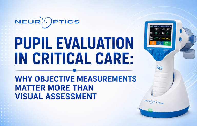
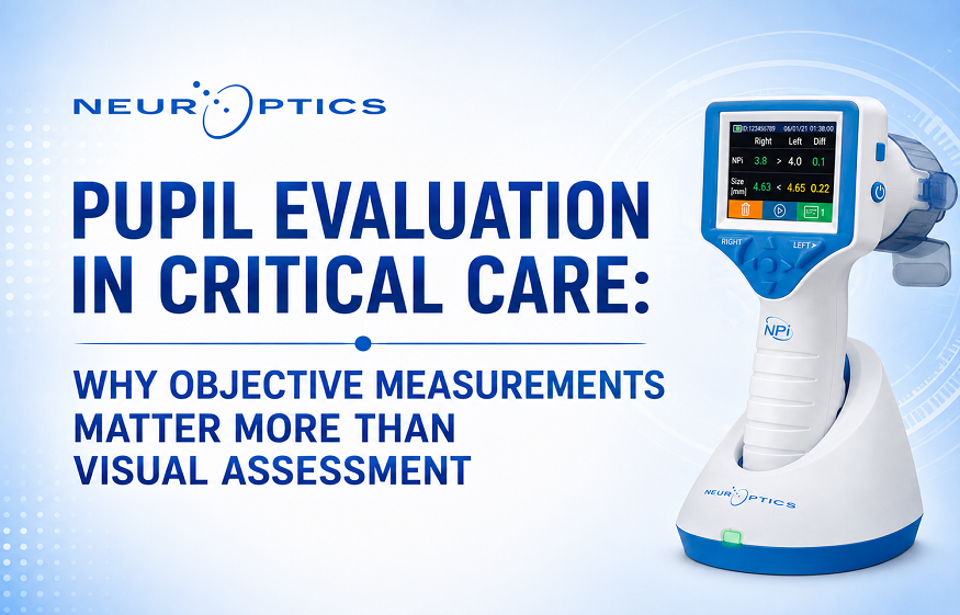
A pupilometer is a handheld infrared device that measures pupil size, constriction velocity, latency, and percent constriction after a fixed light stimulus, converting the readings into one reproducible score. The best known of these is the NPi®, on a 0 to 5 scale, with values under 3 flagged abnormal. Because the device delivers the same light intensity and duration each time, two clinicians checking the same eye get the same number, something a penlight exam cannot promise. It can also shift before pupil size does, giving an earlier warning of change. For pupillary response in traumatic brain injury, trending pupil reactivity score every few hours can flag deterioration before it shows on exam, buying the care team time to act.
Manual Exam vs Automated Pupillometry at a Glance
| Factor | Manual (Penlight) Assessment | Automated Pupillometry |
| Measurement basis | Examiner’s visual judgment | Infrared sensor, standardized light stimulus |
| Interrater reliability | Fair to moderate, kappa 0.40 to 0.62 | High and device-standardized |
| Recorded as | Descriptive terms such as brisk or sluggish | Numeric pupil reactivity score plus size and velocity |
| Time per eye | Varies by examiner | Under 30 seconds |
| Best suited for | Quick bedside screening | Trending and prognostic tracking |
How Should Hospitals Build Objective Pupil Evaluation into Their Neuro Examination Protocols
A pupillometer works best as a partner to the hands-on exam, not a replacement. Steps neuro ICU leaders use to close the reliability gap:
- Pair manual checks with a pupillometer reading during the first 24 to 48 hours after admission for TBI, stroke, or subarachnoid hemorrhage.
- Chart NPi trends over successive shifts instead of one read.
- Set a clear escalation threshold, such as if the score drops below 3, so the number triggers action.
Hospitals that build this into their neuro exam workflow tend to catch pupillary changes hours before visual assessment alone would show them.
Conclusion
Pupil assessment has always been part of the neurological exam, but the neurological toolsused to perform it have not kept pace with how much rides on the result. Manual assessment still has a place at the bedside, yet the interrater data make a clear case for an added objective measurement layer wherever pupil reactivity guides a clinical decision, giving critical care teams a fuller, faster picture of a patient’s neurological status.
Hospital and health system teams looking to standardize pupil assessment across their ICU and emergency departments can connect with NeurOptics, developer of the NPi® Pupillometer system, to see how objective pupillary monitoring fits into existing neuro exam workflows.
Frequently Asked Questions
Q: What does a low NPi score mean for a patient?
Below 3 is considered abnormal and signals reduced pupillary reactivity, which clinicians weigh alongside other neurological findings to assess for rising intracranial pressure. A downward trend across readings carries more weight than any single number.
Q: Can pupillometry replace the neurological exam entirely?
No. Pupillometry adds an objective, reproducible measurement to the exam; it does not replace motor response checks, GCS scoring, or clinical judgment. Most neurocritical care protocols use it alongside the full neuro assessment.
Read More :Why does depression make you tired?
Q: How often should ICU teams check pupil reactivity scorein traumatic brain injury patients?
Frequency depends on acuity and unit protocol, but many neuro ICUs check every 1 to 2 hours during the acute phase after TBI, then space out readings as the patient stabilizes.
About the Author